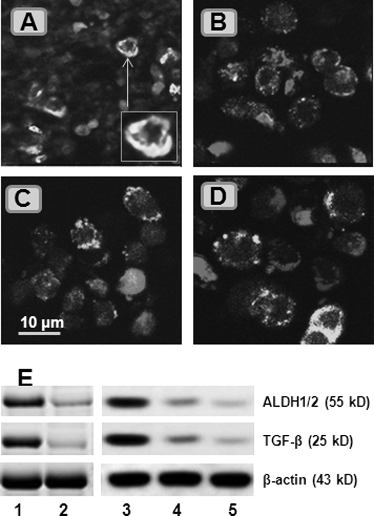
FIGURE 4.

Glioma-derived Mϕs phagocytose apoptotic T cells to become tolerogenic. A, surgically removed glioma tissue was processed for cryosectioning and stained with FITC-conjugated anti-CD14 (1:200) and Cy5-conjugated anti-CD3 (1:100) antibodies and phycoerythrin-conjugated annexin V. The representative confocal image shows CD14-positive staining (in green) and CD3-positive staining (in red). Some CD3+ cells were localized inside CD14+ cells (inset). B–D, CD14+ and CD3+ cells were isolated from peripheral blood mononuclear cells, cultured under hypoxia (B and C) or normoxia (D) for 48 h, and processed for staining with the reagents used in A. The representative confocal images show that some CD14+ cells phagocytosed apoptotic CD3+ cells (labeled with asterisks). E, total proteins were extracted from CD14+ cells (isolated from glioma tissue or peripheral blood) in the glioma group (lane 1), non-glioma group (lane 2), cells in B (lane 3), cells in C (lane 4), and cells in D (lane 5). The immunoblots show the levels of aldehyde dehydrogenase-1/2 (ALDH1/2) and TGF-β. Data represent five experiments. The image original magnification was ×630.
