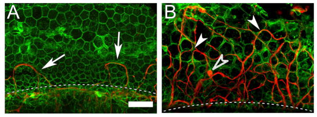
Figure 2. Whole mount of a flat epithelium of a guinea pig cochlea with (B) or withoutAd.BDNFtreatment (A), stained for actin (phalloidin, green) and neurofilaments (red).
The flat epithelium, in the area where the organ of Corti used to reside, is composed of polymorphic cuboidal cells. The intercellular adherens junctions are actin rich. Nerve fibers are mostly absent, except for a few looping fibers (arrow) next to the habenula perforata (dashed line) (A). In a cochlea with flat epithelium treated with Ad.BDNFa large number of re-grown peripheral nerve fibers is present. The fibers appear to traverse the epithelium between the cuboidal cells (arrowheads). Fibers are of different diameter and their orientation in the flat epithelium varies from longitudinal to radial. Some bulging regions that resemble terminals are seen (open arrowhead) (B). Scale bar = 50 μm, for (A) and (B).

