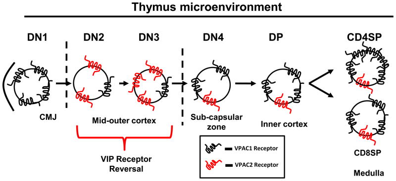
Figure 6. Hypothetical model for VIP receptor axis in the thymus.
Once recruited to the CMJ, ETP cells show exclusive, high VPAC1 levels, which are downregulated upon DN2 differentiation. DN2 and DN3 cells show a coordinate upregulation of VPAC2 expression. VIP receptor levels are restored during later differentiation stages, with VPAC1 levels predominating.

