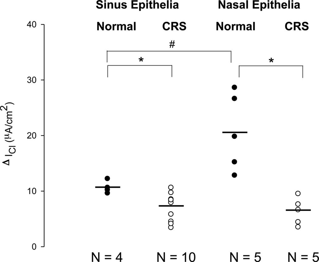
Figure 3.
Changes in basal Cl currents after stimulation with L-ascorbate (ΔICl) in freshly excised sinus and nasal epithelial tissue from normal subjects (Normal) and patients with chronic rhinosinusitis (CRS). L-ascorbate stimulated Cl currents are plotted from individual experiments and average values are shown as horizontal bars. The largest Cl secretory response to L-ascorbate was noted in nasal epithelia from normal subjects. Both sinus and nasal epithelia from CRS patients showed lower Cl secretory responses to L-ascorbate compared to their normal counterparts. #: denotes a significant difference between nasal and sinus epithelial tissue from normal subjects (p<0.05). *: denotes a significant difference between sinus epithelia obtained from normal and CRS subjects (p<0.05). ●: Normal epithelia. ○: Diseased epithelia.

