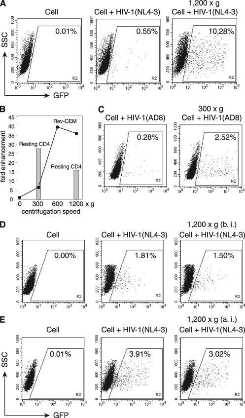
Fig. 1.
Spinoculation enhances HIV-1 infection of transformed and resting CD4 T cells. (A) To demonstrate spin-mediated enhancement of HIV-1 infection, Rev-CEM cells (2 × 10⁵ cells) were not infected (Cell) or were infected with HIV-1NL4-3 for 2 h in the absence or presence of spin at 1,200 × g. SSC, side scatter. (B) Rev-CEM cells were infected similarly at different centrifugal speeds. Shown are the levels of enhancement (fold) based on the percentages of GFP-positive cells spun as shown. Cells were washed twice and cultured for 2 days, and then GFP-positive cells were measured by flow cytometry. Resting CD4 T cells (1 × 10⁶) were also spinoculated at 300 × g or 1,200 × g for 2 h, washed, cultured for 5 days, and then activated with CD3/CD28 stimulation. Shown are the levels of enhancement (fold) of viral replication at day 9 postinfection for cells spun as shown. (C) Rev-CEM cells were infected similarly with HIV-1AD8 at 300 × g. (D) Effects of spinning cells prior to HIV infection. Rev-CEM cells were directly infected with HIV-1NL4-3 or centrifuged at 1,200 × g for 2 h before infection (b. i.) and then infected with HIV-1NL4-3 for 2 h in the absence of spin. (E) Effects of spinning cells after HIV infection. In a separate experiment, Rev-CEM cells were directly infected with HIV-1 for 2 h or infected with HIV-1 for 2 h, washed twice with medium, and then centrifuged at 1,200 × g for 2 h. Cells were cultured for 2 days, and then GFP-positive cells were measured by flow cytometry.

