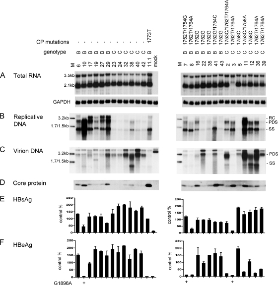
Fig. 5.
Comparison of RNA transcription, genome replication, virion secretion, and viral protein expression/secretion between Chinese genotype B and genotype C isolates. Huh7 cells were transfected with circularized HBV genomes and harvested 2 days later (for RNA analysis) or 5 days later (for other analyses). (Left panels) Isolates lacking core promoter mutations, with genotype B clone 11.1 serving as a control; (right panels) isolates with the core promoter mutations indicated above. (A) Intracellular HBV RNA, with GAPDH serving as loading control. (B) Intracellular replicative DNA. (C) Virion DNA. Lanes M, 3.2-kb, 1.7-kb, and 1.5-kb HBV DNA markers at 100 pg (B) and 10 pg (C). For panels A to C, genotype B and C probes were used and washing was at 62°C with 2× SSC-0.1% SDS. (D) Immunoprecipitation-Western blot analysis of intracellular core protein. (E and F) ELISA for secreted HBsAg and HBeAg based on 3 transfection experiments, with values from clone 11.1 set at 100%.

