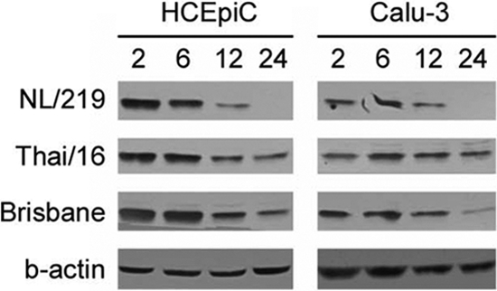
Fig. 4.

Degradation of IκBα following influenza virus infection in human ocular and respiratory cells. HCEpiC or Calu-3 cells were infected with the indicated viruses at an MOI of 2. Protein samples were harvested at 2, 6, 12, or 24 h p.i. and examined by SDS-PAGE for the presence of IκBα. Equal loading was achieved by loading 200 μg of sample per well and was confirmed by β-actin (a representative blot is shown).
