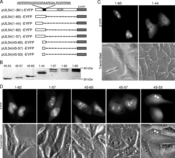
Fig. 2.
The nucleolar localization signal locates in the arginine-rich region of UL54. (A) Schematic representation of the WT UL54 protein and its deletion mutants fused with EYFP; (B) Western blot analysis of the deletion derivatives of UL54 using anti-YFP pAb; (C) subcellular localization of deletion mutants aa1-65-EYFP and aa1-44-EYFP; (D) subcellular localization of the other UL54 mutants fused with EYFP (arrowheads, nucleoli of the cells). Each fluorescence image is representative of the vast majority of the cells observed.

