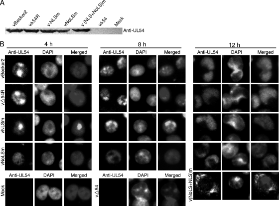
Fig. 6.
Subcellular localization of UL54 in different recombinant virus-infected PK-15 cells during infection. (A) Western blot analysis of different recombinant virus-infected PK-15 cells. Monolayer PK-15 cells were infected with distinct recombinant viruses, including vBecker2, vΔUL54, vΔUL54R, vNLSm, vNoLSm, and v(NLS+NoLS)m viruses, and the cells were collected when CPE reached 90 to 95%. Then the cell lysates were subjected to Western blot analysis using an anti-UL54 pAb. (B) IFA was carried out to characterize the subcellular localization of UL54 in PRV-infected PK-15 cells. PK-15 cells infected with vBecker2, vΔUL54R, vNLSm, or vNoLSm virus were fixed at different time points postinfection (4, 8, and 12 h) with 4% paraformaldehyde, permeabilized with 0.5% Triton X-100, and probed with anti-UL54 pAb. For v(NLS+NoLS)m virus, vΔUL54 virus, and mock infection, PK-15 cells were examined only at 12 hpi. Cells were treated with FITC-conjugated goat anti-mouse IgG and counterstained with DAPI to visualize the nuclei.

