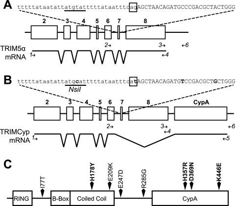
Fig. 1.
Genomic organization and mRNA splicing of TRIM5 alleles. (A) TRIM5α-expressing allele. At the top is the sequence of the intron 6-exon 7 junction, with the intronic sequence in lowercase type and exon 7 in uppercase type. The canonical AG splice acceptor dinucleotide is boxed. The underlined sequence represents the location of the NsiI restriction site in the other allele. Below is a schematic of the TRIM5 gene, with open boxes representing exons 2 to 8, numbered in boldface type. The mRNA splicing pattern is indicated below. (B) TRIMCyp-expressing allele. Sequence changes from the sequence in panel A are in boldface type, and the NsiI site is labeled. Below, the CypA insertion in the TRIMCyp-expressing allele is located as indicated. Minor splice isoforms in both alleles are not depicted. Primers used for analyses described in the subsequent figures and the text are depicted as arrows and numbered in italics. Primers used for genomic analysis are shown above the mRNA diagram, and those used for RT-PCR analysis are shown below. Primer 4 was used for both analyses. (C) Primary structure of TRIMCyp, showing polymorphisms found in M. fascicularis. Residues known to affect protein function are marked in boldface type.

