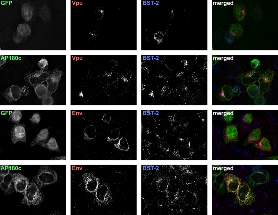
Fig. 5.
The C-terminal fragment of the clathrin assembly cofactor AP180 does not affect the subcellular distribution of Vpu or Env. Cells (HeLa) were transfected to express either the AP180 C terminus fused to GFP (AP180-C; 0.35 μg of plasmid) or GFP (0.03 μg of plasmid), along with either Vpu (0.1 μg of plasmid) or HIV-2 Env with HIV-1 Rev (0.1 μg of each plasmid); total plasmid was made up to 0.8 μg in each case with the empty vector pCDM8. The next day, the cells were fixed, permeabilized, and stained for Vpu or Env, together with BST-2, and imaged using wide-field fluorescence microscopy. A Z series of images was obtained, and these were processed by a deconvolution algorithm before export of the single-plane images shown. In the “merged” images, GFP proteins are shown in green, Vpu or Env is red, and BST-2 is blue. Overlap between the viral proteins and BST-2 appears purple, whereas overall between the viral proteins and AP180-C or GFP appears yellow.

