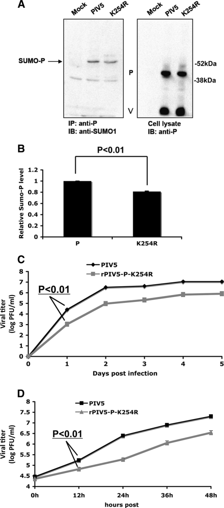
Fig. 4.
Sumoylation of K254R in virus infection. (A) Sumoylation of P-K254R in infected cells. HeLa cells were mock infected or infected with PIV5 or rPIV5-P-K254R at an MOI of 5. At 24 hpi, the cells were lysed and the supernatants used for IP-IB. (B) Quantification of the sumoylation levels. Three individual experiments from panel B were performed for quantification and statistical analysis. (C) Growth rate of rPIV5-P-K254R at an MOI of 0.01. MDBK cells were infected with PIV5 or rPIV5-P-K254R, and the supernatants were collected at different time points for plaque assay. (D) Growth rate of rPIV5-P-K254R at an MOI of 3 in MDBK cells.

