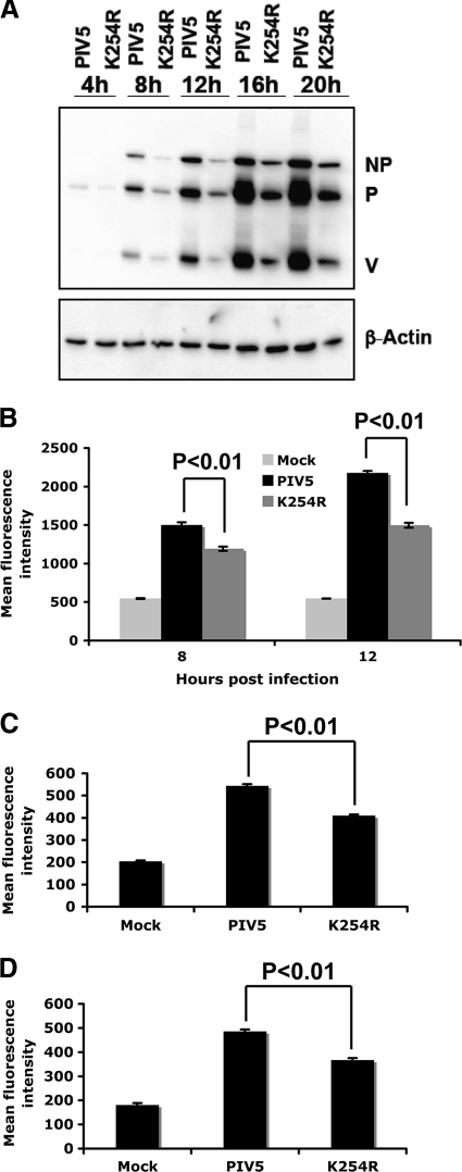
Fig. 5.
Viral protein expression levels in rPIV5-P-K254R-infected cells. (A) Immunoblotting. MDBK cells were infected with PIV5 or rPIV5-P-K254R at an MOI of 3. The cells were collected at different time points and used for immunoblotting using anti-NP and Pk (anti-P/V) antibodies. β-Actin was used as a protein loading control. K254R indicates rPIV5-P-K254R virus. (B) Flow cytometry. MDBK cells were mock infected or infected with PIV5 or rPIV5-P-K254R at an MOI of 3. Flow cytometry was performed to compare viral protein expression at different time points using Pk antibody. (C and D) Flow cytometry in HeLa (C) and BSR-T7 (D) cells. Similar experiments were performed at an MOI of 1 in HeLa and BSR-T7 cells at 10 hpi.

