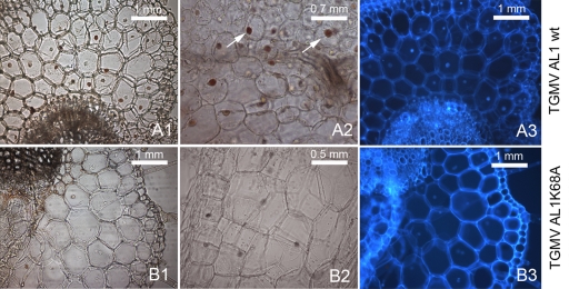
Fig. 5.
Immunolocalization of TGMV in infected N. benthamiana plants. Panels A1, A2, and A3 correspond to wild-type TGMV A- and B-inoculated plants, while panels B1, B2, and B3 correspond to plants infected with TGMV B and the TGMV A K68A mutant. Panels A3 and B3 are the same sections as those in panels A1 and B1, respectively, but with 4′,6-diamidino-2-phenylindole (DAPI)-stained nuclei. Sectioning was performed 19 days after plant infection, and a polyclonal AL1 primary antibody was used. Arrows indicate examples of infected nuclei.

