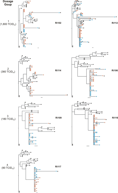
Fig. 4.
ML trees of envelope sequences amplified from samples obtained at week 2 and week 12 postchallenge along with sequences from the inoculum. Red squares, week 2 animal-derived sequences; blue squares, week 12 animal-derived sequences; black circles, inoculum-derived sequences. Bars, 1 nucleotide substitution. Asterisks indicate hypermutated sequences.

