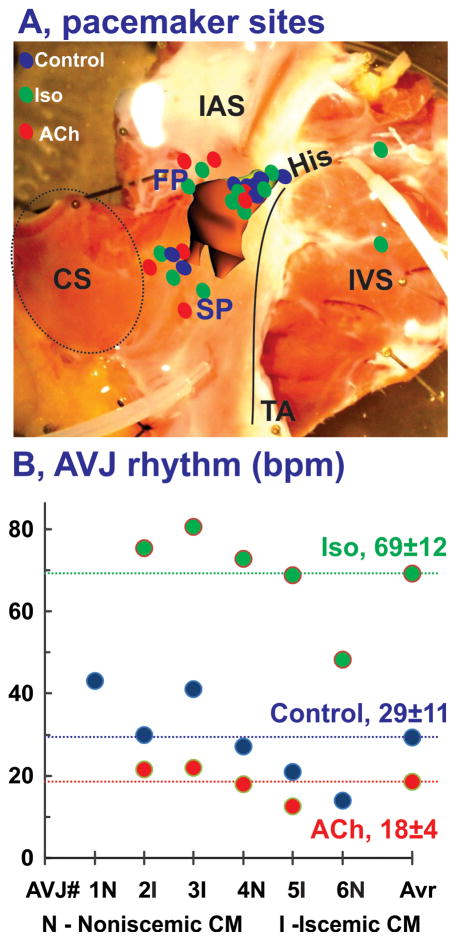
Figure 7.
Summary of anatomic locations of leading pacemaker sites in the human AVJ. Panel A: Summary of pacemaker sites identified in all hearts (n=6) in control (blue), under 1 μM Iso (green), and 1 μM ACh (red). Panel B: Individual for each heart and average (Avr) values for AVJ rhythm in control and during perfusion of Iso and ACh. CM- cardiomyopathy.

