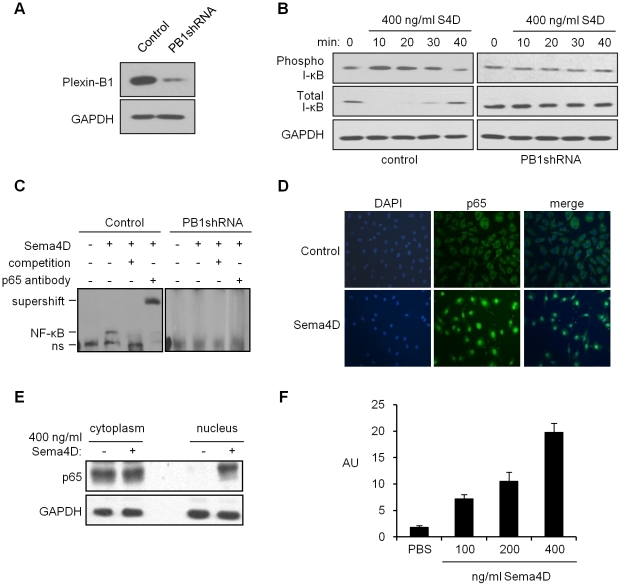
Figure 1. Sema4D activates NF-κB downstream of Plexin-B1.
A. Immunoblot analysis for Plexin-B1 on lysates from endothelial cells infected with empty vector control lentivirus (control) or virus coding for Plexin-B1 shRNA (PB1shRNA). GAPDH was used as a loading control (lower panel). B. Immunoblot for phospho- and total I-κB (upper and middle panels, respectively) in endothelial cells infected with empty vector control lentivirus (control, left column) or virus coding for Plexin-B1 shRNA (PB1shRNA, right column) treated with Sema4D for the times indicated. GAPDH was used as a loading control (lower panels). C. EMSA performed on control infected HUVEC or cells infected with lentivirus coding for Plexin-B1 shRNA, in the presence of Sema4D with and without competition from unlabeled oligos (competition) or anti-p65 antibody (p65 antibody). “ns” denotes a non-specific band; “NF-κB” indicates a shift in oligo migration; “supershift” indicates the higher band resulting from antibody binding. D. Immunofluorescence for the presence of the p65 subunit of NF-κB in nuclei of HUVEC, controls (top row) or treated with Sema4D (bottom row). p65 is shown in green (center column). DAPI was used to stain cell nuclei (left column). The merged image is shown in the right column. E. Immunoblot for the presence of p65 in cytoplasmic and nuclear fractions of HUVEC treated with Sema4D. GAPDH was used as a loading control (lower panel). F. HUVEC were infected with lentiviruses coding for an NF-κB reporter construct and fluorescence measured in arbitrary units (AU) in increasing concentrations of Sema4D, as indicated. Error bars represent the standard deviation from three experiments.

