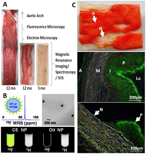
Figure 1. Rabbit aorta cholesterol plaque formation and NP diffusion into intima in vivo.
A: Sudan IV staining (en face) of opened aorta section showing plaque (red) in 12 month diet rabbit but seldom in 3 month diet rabbit. Sections were collected as labeled for fluorescence microscopy and histology (1 cm), electron microscopy (0.5 cm), lower segments for MRI/MRS and whole mount fluorescence images (IVIS). B: Cartoon of PFC-NP structure, and 19F MR spectroscopy (top left), TEM of NP (top right); 19F and 1H MR image of test tubes containing CE core NP showing fluorine signatures (bottom) and oil core NP as control showing no signal. C: Top: En face Sudan IV staining of section with plaque (P: red) and grossly normal (N: clear) areas. Mid: Marked fluorescent nanoparticle presence in plaque (P) intima (I) of 12 mo cholesterol diet rabbit aorta (green) after 12 hours in vivo circulation. Minimal staining of adventitia (A) is noted , and none apparent in media (M) or lumen (Lu). Bottom: Fluorescent NP signals (green) in plaque intima (P), but not the adjacent grossly normal regions (N). Blue = DAPI nuclear stain.

