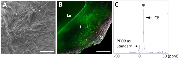
Figure 6. SEM, fluorescence microscopy and MRS of human carotid endarterectomy specimen.
A: SEM image of intimal subendothelial cholesterol crystals in carotid endarterectomy tissue. (scale bar = 50 um) B: Fluorescence microscopy of human carotid endarterectomy specimen (fresh) incubated ex vivo with Alexa Fluor-488 labeled NP (green). Lu: lumen; I: intima; M: media. (scale bar = 500 um) C: 19F MR spectroscopy of human plaque segment indicating a strong signal and the presence of trapped CE NP in plaque. (ppm: part per million). PFOB standard signal from co-registered control sample is for NP calibration.

