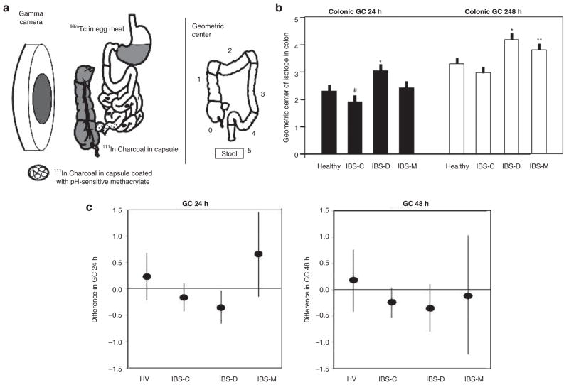
Figure 1.
Colonic transit measurement by scintigraphy in health and subgroups of IBS. (a) Method to measure colonic transit: radioisotope-labeled activated charcoal particles delivered in a pH-sensitive methacrylate-coated capsule to the ileocolonic region. Methacrylate dissolves at pH >6.4, which is achieved in the distal small intestine. The radiolabeled particles traverse the colon in the stool, and the counts in each of five regions are used to summarize the location of the weighted average or geometric center at specified times after capsule ingestion, e.g., at 24 and 48 h. More frequent imaging allows accurate estimation of the ascending colon emptying time. The ascending colon emptying time is significantly correlated with stool consistency. (b) Comparison of colonic geometric center at 24 and 48 h in healthy controls and in IBS patients with different types of bowel dysfunction. #P = 0.078; *P < 0.05; **P = 0.056. Reproduced from ref. 6. (c) Changes in geometric center in various subgroups expressed as means and 95% confidence intervals. Note that the 95% confidence interval for the IBS-D subgroup does not cross the zero line, indicating a significant difference in colonic transit time for the IBS-D subgroup but not for the other groups. Reproduced from ref. 9. GC, geometric center; HV, healthy volunteers; IBS, irritable bowel syndrome; IBS-C, constipation-predominant IBS; IBS-D, diarrhea-predominant IBS; IBS-M, IBS with mixed bowel function.

