Fig. 3.

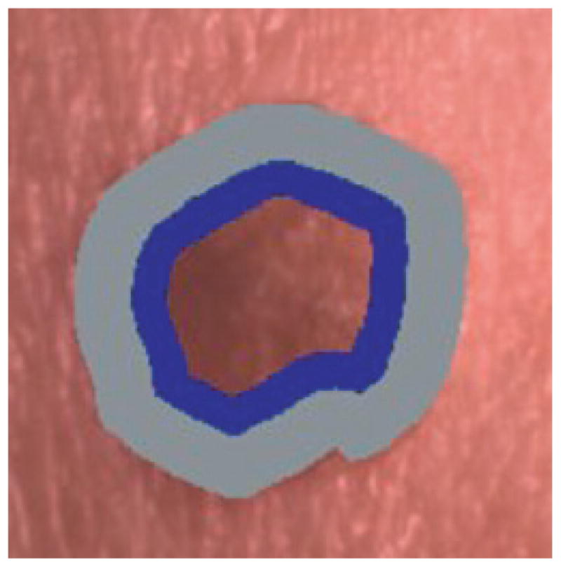
Offset boundary area example using 50% of the lesion area as offset (gray region) and 25% of the lesion area for analysis (black region). (This is the same lesion as in Fig. 1(b)).

Offset boundary area example using 50% of the lesion area as offset (gray region) and 25% of the lesion area for analysis (black region). (This is the same lesion as in Fig. 1(b)).