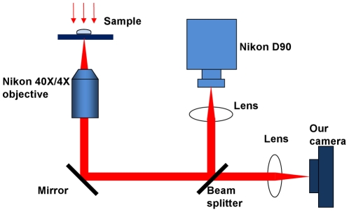
Figure 10. Experimental setup.
A live Daphina (water flea) is placed in a small chamber on the stage of a microscope (Nikon ECLIPSE Ti). A commercial camera (Nikon D90) is used to obtain high pixel-count wide-field images while our camera is used for high-speed observation. Different objectives (4×, NA = 0.13 and 40×, NA = 0.74 respectively) are used to observe the beating heart and the flowing blood cells of the Daphnia.

