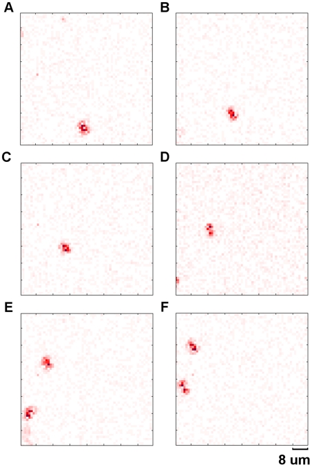
Figure 15. Blood cell flow.
Sequence of differential frames showing individual cells flowing along blood vessels in a live Daphnia. These images were acquired in full frame mode at 24.6 fps. Frame 1 (A), 8 (B), 15(C), 22(D), 29(E) and 36(F). These differential images are obtained by subtracting adjacent frames. A second blood cell can be seen entering into the field of view in frame 22 (D) (see supplementary material Movie S4).

