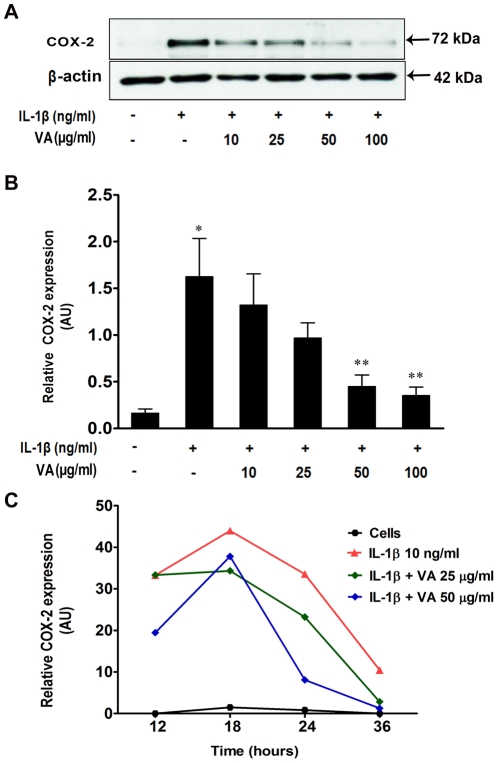
Figure 3. Inhibition of cytokine-induced COX-2 protein expression by Viscum album in A549 cells.
A549 cells were either cultured in medium alone or treated with IL-1β (10 ng/ml) with or without increasing concentration of VA Qu Spez for 18 hours. Using 20 µg of total cellular protein, expression of COX-2 was analyzed by western blotting. A. A representative western-blot depicting the effect of VA on expression of IL-1β-induced COX-2. B. Relative expression (mean ± SEM) of COX-2 protein from four independent experiments as quantified by densitometry (ratio of density of COX-2: that of β-actin). *P<0.05 versus control cells and ** p<0.05 versus IL-1β-stimulated cells as analyzed by paired Student-t-test. C. Kinetics of COX-2 protein expression upon treatment of A549 cells with various doses of VA. The cells were either cultured in medium alone or stimulated with IL-1β with our without VA for 12, 18, 24 and 36 hours. Expression of COX-2 protein was analyzed by western blotting and relative expression of COX-2 protein was quantified by densitometry (ratio of density of COX-2: that of β-actin). Results are representative of two experiments.

