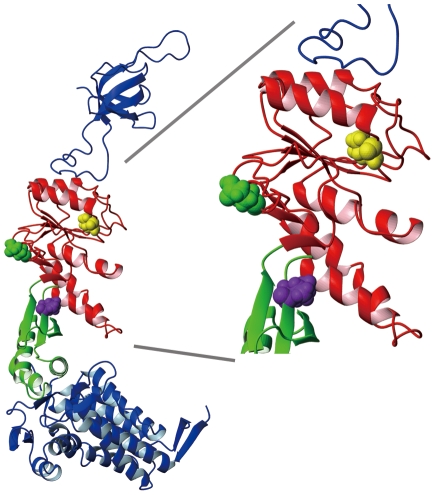
Figure 1. Locations of the N- and C-termini in the three circular permutation mutants of the present study.
An X-ray structural model of a single GroEL/GroES subunit pair taken from PDB structure file 1 AON [3], depicting the three locations where the N- and C-termini were relocated in the three CP mutants. The locations are indicated by colored CPK representation of the first (N-terminal end) amino acid of each CP mutant, excepting the starting methionine and any extraneous amino acids that were added as a consequence of the experimental protocol (see Materials and Methods). The green molecule denotes Glu 209, the yellow molecule Val 254, and the magenta molecule Val 376 in wild type GroEL.

