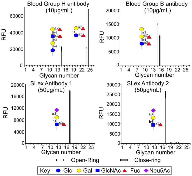
Fig. 3.
Comparison of 4 different antibodies binding to open-ring and closed-ring AEAB conjugates. The arrays were printed using a piezo printer (Perkin Elmer) with open- and closed-ring AEAB derivatives at 300 μM on NHS-derivatized slides. Antibodies were applied to the glycan array at the indicated concentrations above and detected with appropriate fluorescently labeled secondary antibodies (Song et al., 2009). The X-axis represents different glycans and the Y-axis represents the relative fluorescent unit (RFU) detected on the microarray.

