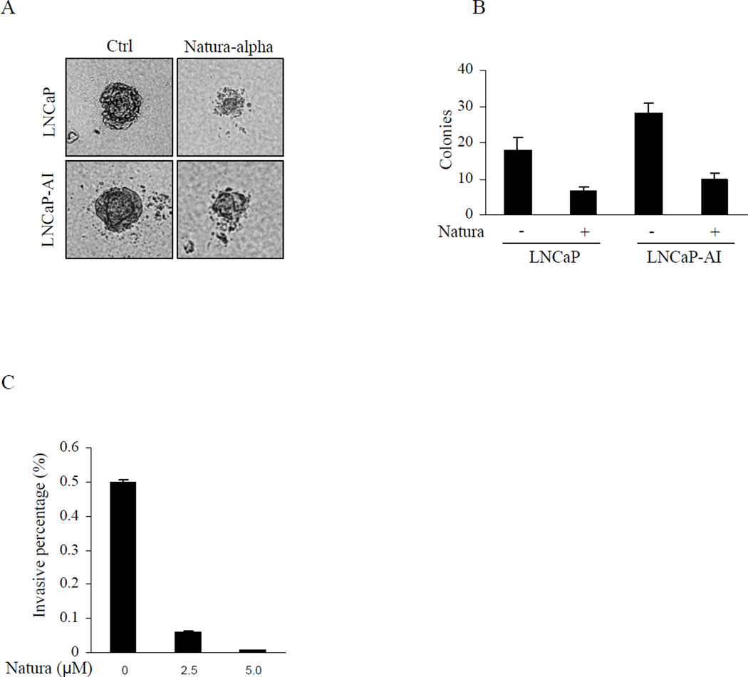
Figure 2.
Inhibition of prostate cancer anchorage independent growth and invasion. A and B: Anchorage independent cell growth in soft agar was performed in triplicate with cells (1 × 10⁴) suspended in 2 mL of medium containing 0.35% agar (Becton Dickinson) spread on top of 5 mL of 0.7% solidified agar. Colony volume was calculated from the average radius of representative colonies. C: LNCaP-AI cells at their exponential growth phases were added to the upper chamber at density of 1×10⁴ cells in 500ul medium in the presence or absence of indicated concentrations of Natura-alpha and incubated at 37° C for 48 hrs, and the invading cells adherent at the bottom of the membrane were fixed, stained, and counted by tallying the number of cells in 3 random fields under the microscope. Data were adjusted by growth condition, and expressed as mean of migrating cells in 3 fields ±SD.

