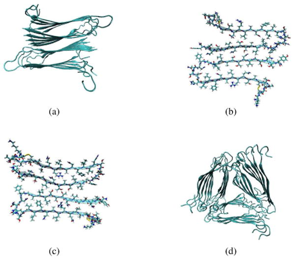
Fig. 2.

(a) The overall backbone arrangement of the two-fold protofilament structural models.7 (b) The cross section of the two-fold model I, which corresponds to the structure depicted in Figure 11C of reference 7. (c) The cross section of the two-fold model II, which corresponds to the structure depicted in Figure 11D of reference 7. (d) The overall backbone arrangement of the three-fold protofilament model.92 In each diagram, for each peptide the inner strand ends with the C-terminus (and the outer strand ends with the N-terminus).
