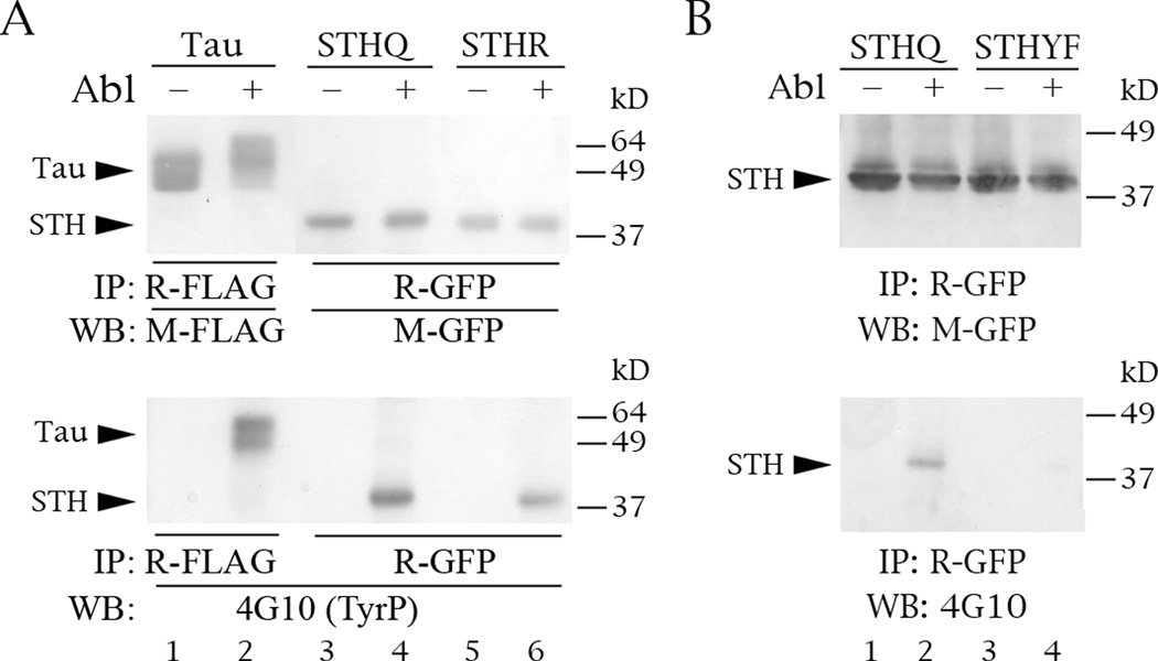
Fig. 5.
Abl phosphorylates STH on its single tyrosine. The Western blots are of protein lysate inputs (top and middle panels) and IPs (bottom panels) from co-transfections into COS cells. (A) Phosphorylation of FLAG-tau, GFP-STHQ and GFP-STHR by Abl. (B) Abl does not phosphorylate mutant STHYF, in which tyrosine 78 has been changed into a phenylalanine. Labeling conventions as the same as in Fig. 3.

