
Fig. 6.
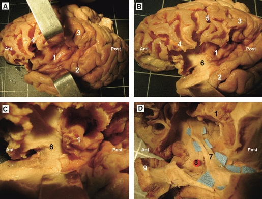
Stepwise dissection of the UF and the IFOF in a left hemisphere. (A) The lateral surface of the temporal, occipital, parietal and frontal lobes has been dissected, enabling us to lift the opercula, allowing the removal of the hidden cortex of the inner surface of the operculum, exposing the insular cortex. (B) The antero-inferior portion of the insular cortex has been removed, exposing the extreme and external capsule. (C) Enlarged view of the previous picture, where the curving fibers of the UF are observed. (D) Further dissection of the antero-inferior portion of the insular lobe, at this region the UF and the IFOF narrow in section and the IFOF is located dorsal and posterior to the UF. 1, insular lobe; 2, superior temporal gyrus; 3, inferior parietal lobe; 4, frontal operculum; 5, central sulcus; 6, antero-inferior portion of the extreme and external capsule; 7, IFOF; 8, UF; 9, temporal pole. Ant, anterior; Post, posterior.