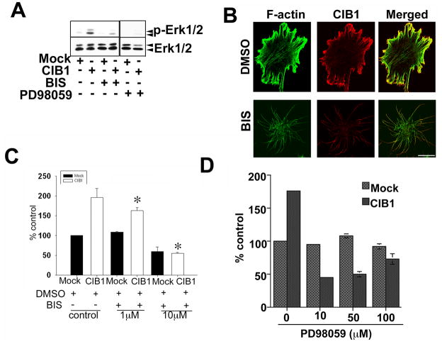
Figure 3.
CIB1-induced cell migration inhibited by PKC and MEK inhibitors. A: Western blot analysis of cell lysates from mock or CIB1- overexpressing cells treated with DMSO or with 10 μM inhibitors. Blots were incubated with phospho-specific anti-Erk1/2 (p-Erk1/2) and reprobed for total Erk1/2 to ensure equal loading of proteins in the lanes. Shown is a representative blot from three separate experiments. B: Confocal images of serum-starved cells treated with DMSO or 10 μM PKC inhibitor for 20 min. CIB1 localization at the tip of the filopodia and intact actin-fibers (upper panel), treatment with the bisindolylmaleimide I, a protein kinase C inhibitor (BIS) (lower panel; scale bar 10 μm). C&D: Quantitative analysis of transwell migration assay of mock or CIB1-overexpressing cells untreated or treated with BIS or PD98059, MEK inhibitor and allowed to migrate on Fn for 5 h. Triplicate inserts were used for each treatment. Ten random views of each insert were counted. Data is expressed as percent control. Number of mock cells migrating across the membrane was considered 100 percent. Each experiment was repeated independently three times. *P<0.05 vs untreated CIB1 overexpressed (control).

