
Figure 6.
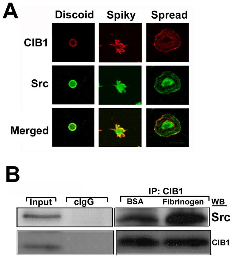
CIB1 and Src associate when platelets spread on immobilized Fg. A: Confocal images of washed platelets at various stages of spreading on immobilized Fg. Shown are the representative images of three different progressive stages of platelet spreading on immobilized Fg. Scale bar 20μm. B: Western blot analysis of platelet lysates exposed to BSA or attached on immobilized Fg. CIB1 was immunoprecipitated and the immunocomplex was subjected to Western blotting. The above membrane was probed with anti-Src (upper panel), and again reprobed with anti-CIB1 to ensure equal loading (lower panel).