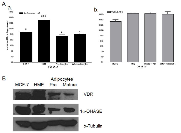
Figure 1. Expression of Vitamin D3 signaling components in human primary breast adipocytes.

A) Real time PCR for 1α-hydroxylase (1α-OHase) (a) and vitamin D3 receptor (VDR) (b) in MCF-7 human breast cancer cells, non-transformed human mammary epithelial cells (HME), and adipocytes (pre and mature adipocytes). Data are expressed relative to 18S RNA (normalized gene equivalence) and represent mean s.e.m. of triplicate runs. Similar letters above the bars designate significance in the expression of 1α-OHase between cell lines- p<0.05. B) Western blot of VDR and 1α-OHase in MCF-7, HME and adipocytes compared to tubulin, which was used as a loading control.
