
Figure 2. Localization of VDR and 1α-OHase in human primary breast adipocytes.
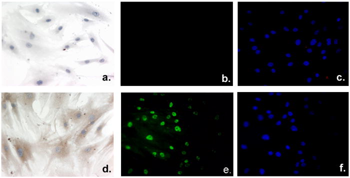
Adipocytes were plated and grown on chamber slides, formalin fixed, and either processed as unstained control cells (a and b) or stained to detect 1α-OHase (d), VDR (e), or the nuclear compartment using DAPI incorporation (c and f). Primary adipocytes were incubated with 1α-OHase (d) or without (a) primary antibody directed against 1α-OHase protein to assess the expression and localization within primary adipocytes. 1α-OHase expression is shown as brown cytoplasmic staining (d) against the blue hematoxylin counterstain (a and d). Immunofluorescent detection of VDR (e) shows nuclear staining within the adipocytes compared to the no primary antibody control (b). Nuclear incorporation of DAPI staining (c and f) of control cells (b) or VDR stained cells (f) emphasizes the nuclear localization of VDR expression.