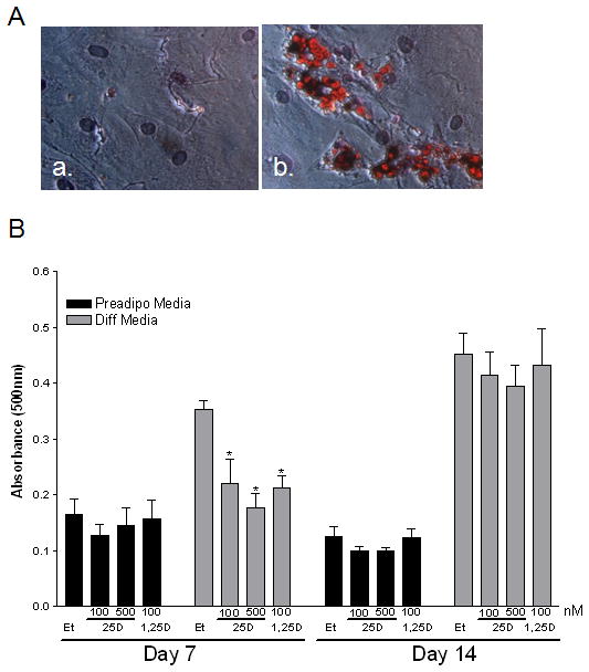
Figure 4. Differentiation of human primary breast preadipocytes in the presence and absence of vitamin D3.

A) Representative image (320x) of preadipocytes (a) and mature (b) adipocytes stained with Oil red O after 14 days of culture in preadipocyte media (a) or differentiation media (b). Oil red O was used to assess intracellular triglyceride and measure lipid content within the cells. The lipid droplets appear red/orange in a halo shape around the nuclei of mature adipocytes. B) Effect of vitamin D3 to influence preadipocyte differentiation in the presence of preadipocyte media or differentiation media. Cells were exposed to vehicle or varying concentrations of vitamin D3 for 7 (a) or 14 (b) days. Assays were terminated on Day 7 or 14 and stained with Oil red O, dried, eluted from the plate with 100% isopropanol to quantitate the absorbance at 500nm in response to vitamin D3 in the preadipocyte or differentiation media. *- p <0.05 compared to vehicle control at each time point.
