
Figure 6. Preadipocytes contribute to vitamin D3-induced growth inhibition of mammary organoids.
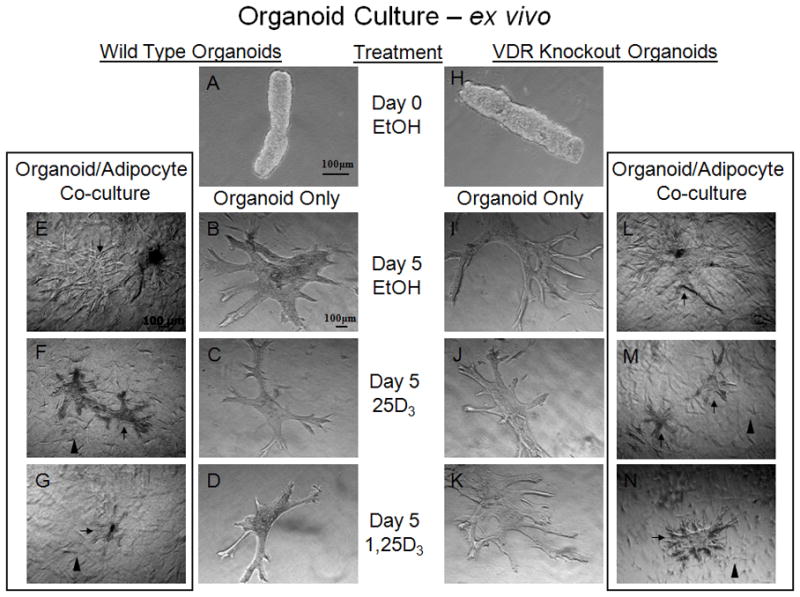
VDR WT (A–G) and KO (H–N) organoids (isolated mammary ductal structures, cleared of surrounding adipose) cultured in Type I Collagen in the presence of vehicle (ethanol) (Day 0 (A & H) or Day 5 (B & I)), 25D3 (100nM) (C & J), 1,25D3 (100nM) (D & K). WT organoids (C) show a slight reduction in branching with 25D3 (100nM) treatment, suggesting bioactivation of vitamin D3 via the mammary ductal epithelial cells compared to KO organoids (J) that show no reduction in size of branching compared with the ethanol control groups (B & I, respectfully). Further reduction in WT organoid (D) size and branching was seen with 1,25D3 treatment compared to KO organoid (K) and ethanol control groups (B & I). VDR wild type adipocytes (arrowheads, visible in F,G,M,N) cultured with VDR WT (E–G) or KO (L–N) organoids (arrows) embedded in Type I Collagen in the presence of vehicle (ethanol) (Day 5 (E & L)), 25D3 (100nM) (F & M), 1,25D3 (100nM) (G & N). Note the reduction in WT and KO organoid size and ductal branching when cultured with wild type adipocytes in the presence of 25D3 (100nM) (F & M) compared with the ethanol control groups (E & L) and without adipocyte co-culture (C & J), suggesting bioactivation via wild type adipocytes. There was also a similar reduction in WT and KO organoid growth when treated with 1,25D3 (G & N), suggesting VDR signaling and growth inhibitory secretion via WT adipocytes that results in growth inhibition of VDR KO organoids. n=3–5 organoids/genotype/treatment and n>5 branches measured/organoid.