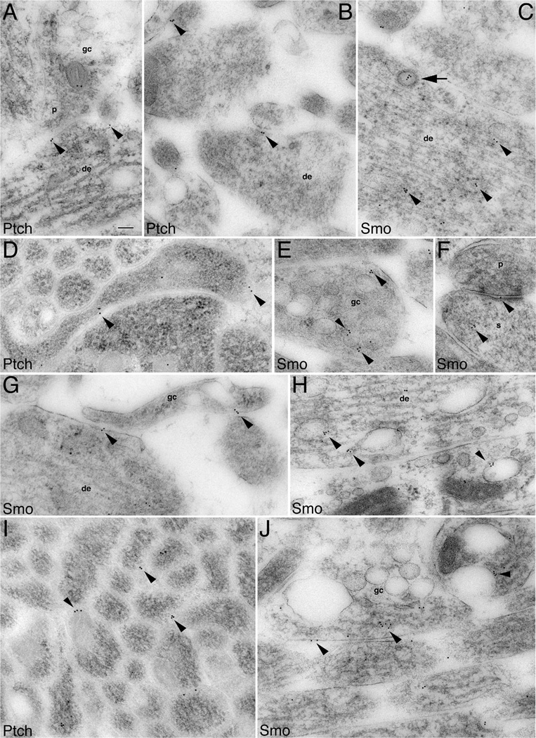
Figure 3.
Subcellular localization of Ptch and Smo in immature processes and growth cones of developing hippocampal neurons. Immunogold labeling for Ptch and Smo in P2 hippocampus CA1 stratum radiatum (A,F,H), and the developing white matter plus stratum oriens dorsal to the CA1 stratum pyramidale (B–E,G,I,J). Ptch labeling: A: An axonal growth cone (gc) forms an immature presynaptic terminal (p) contacting a young dendrite (de) where Ptch labeling is located at the membrane of the contact points (arrowheads). B: Additional examples of Ptch labeling situated at the contacts (arrowheads). D: An unidentified growing process in which Ptch labeling is at contacts with adjacent processes. I: Ptch labeling located in between axons of a large axon bundle (transverse view). Smo labeling: C: A presumptive newly generated neuron with abundant microtubules and neurofilaments shows Smo labeling, which associates with cytoplasmic organelles (arrowheads) including a clathrin-coated vesicle (arrow). E: Smo labeling is found near or on endosomes (arrowheads) that are highly abundant in growth cones. F: Smo labeling in the spine(s) of a nascent synapse. G: Smo labeling is seen at the contacts of a growth cone with a young dendrite and an unidentified process. H: Smo labeling on endosomes of young dendrites. J: Longitudinal view of axons: one of these axons is expanded into a large growth cone. Smo labeling (arrowheads) is seen at the plasma membrane and near or on endosomes. Scale bar = 100 nm in A (applies to A–J).

