Figure 8.
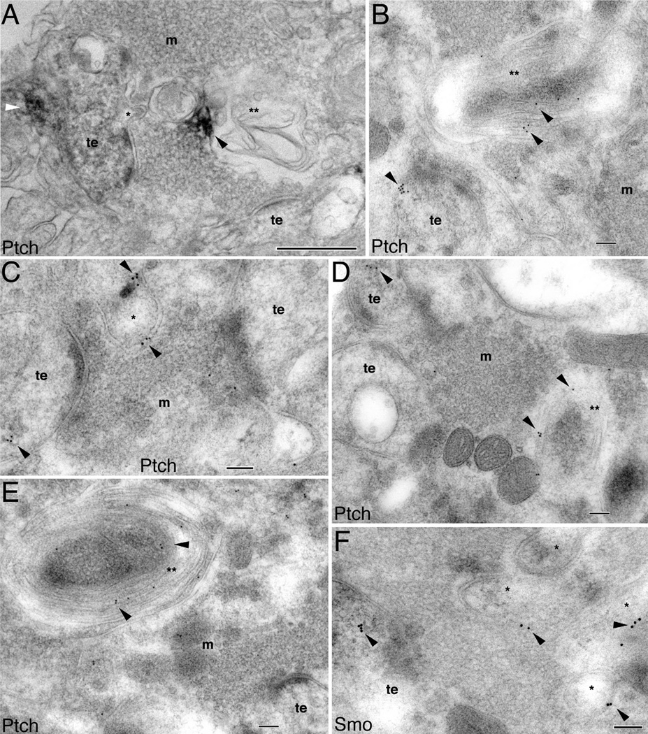
Association of Ptch and Smo with synaptic spinules and autophagosomes in the CA3 mossy terminal area of the adult hippocampus. A is immunoperoxidase/DAB labeling and B–F are immunogold labeling. A: A Ptch-labeled thorny excrescence (te) with a spinule (*) that is oriented toward the surrounding mossy terminal (m); Ptch labeling (white arrowhead) is not within the spinule. Ptch labeling (black arrowhead) is also seen in a nearby irregular shaped autophagosome (**). Note the near contiguous sequence of three structures in the mossy terminal, from the spinule, to a larger spinule-like structure, to the even larger, labeled autophagosome. B: Ptch immunogold labeling (arrowheads) in a thorny excrescence (te) and in a nearby large autophagosome (**). C,D: Ptch immunogold (arrowheads) within the thorny excrescences (te), a relatively large spinule (*), and autophagosome (**). E: Ptch immunogold bonded to the plasma membranes of autophagosome whorls (**). F: Examples of Smo immunogold in the large spinules (*) in addition to the thorny excrescence (te). Scale bar = 500 nm in A; 100 nm in B–F.

