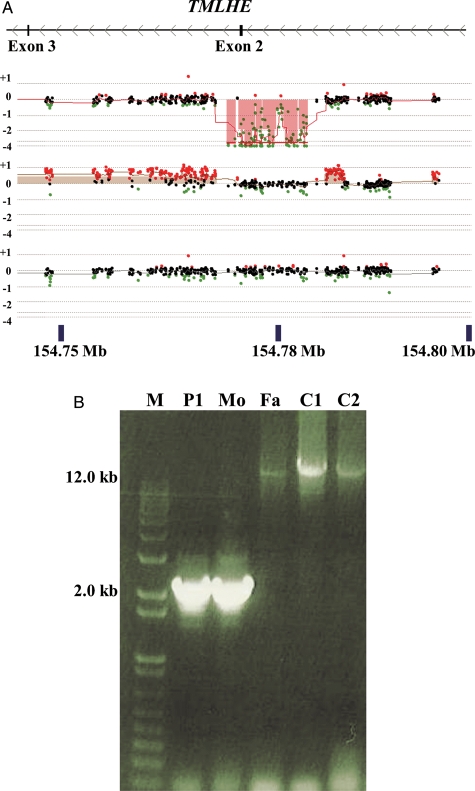
Figure 4.
Validation of hemizygous deletion of exon 2 of TMLHE. (A) The browser display and labels are as in Fig. 2 showing exons 2 and 3 of TMLHE (UCSC genome browser GRCh37/hg19 assembly). Analysis of family proband SSC 11000 (top), mother (middle) and father (bottom) is shown. (B) PCR for the SSC 11000 family showing the deletion in the patient (P1) and mother (Mo), but not in the father (Fa) or unaffected controls (C1 and C2). There is bias of amplification of the smaller band in the mother so that the normal band is faint.

