
Figure 1.
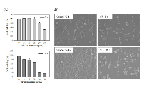
Cell viability of TM4 cells treated with NP and phase contrast microscope images. (A) Measurement of viability using CCK-8. All experiments consisted of three independent replicates, each performed at least in triplicate. Data are presented as the mean ± SD. (B) Morphological observation of NP-treated TM4 cells. TM4 cells were treated with 10 μg/ml NP.