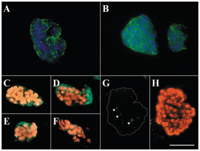
Figure 2.
Maintenance of cell–cell junctions and basement membrane components in HCEC aggregates. Immunofluorescence staining showed ZO-1 (A), connexin-43 (B), type IV collagen α1 (C) and α2 (D) chains, laminin 5 (E), and perlecan (F) were present in HCEC aggregates. The TUNEL assay revealed only a few apoptotic cells present in the center of the aggregate (G, arrowheads; H shows the nuclear counterstaining of G). Bar, 100 μm.

