Figure 5.

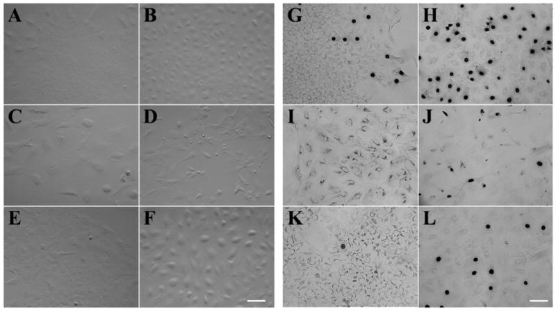
Ki67 expression of HCECs cultured under different conditions. HCEC aggregates harvested from two donors, 48 and 53 years old, without (A, C, E) or with (B, D, F) a brief trypsin/EDTA treatment were seeded on plastic in SHEM (A, B), SHEM+BPE (C, D), or SHEM+NGF (E, F) for 1 week. Immunostaining showed sporadic Ki67-positive nuclei in the periphery of HCEC sheets when aggregates were cultured in SHEM (G). In contrast, very few Ki67-positive nuclei were found in SHEM+BPE (I) and SHEM+NGF (K). Ki67-positive nuclei increased dramatically when aggregates were pretreated with trypsin/EDTA (H, J, L). However, there were more Ki67-positive nuclei in SHEM (H) than those in SHEM+BPE (J) and SHEM+NGF (L). Bar, 100 μm.
