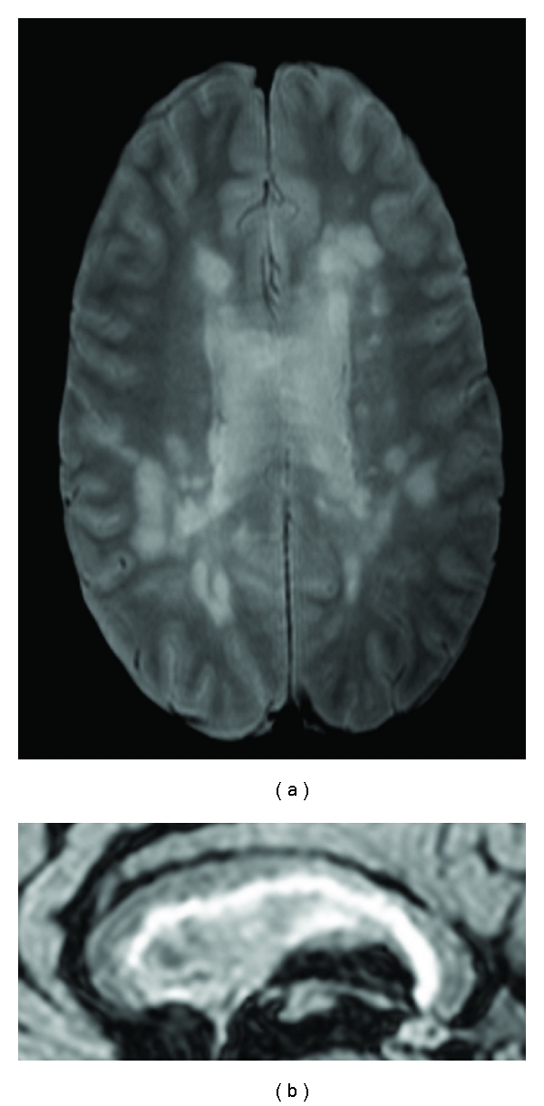
Figure 2.

Female patient with high lesion load and no significant cognitive deficit. The PD axial image at the level of the body of the lateral ventricles and CC (a) shows lesions at the periventricular and subcortical white matter. The detail of mid-sagital FLAIR (b) shows the abundance of macroscopic lesion in the CC and callosal-septal interface.
