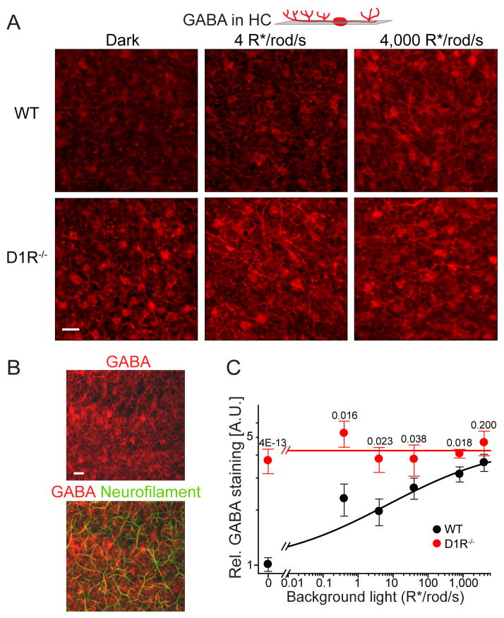
Figure 8. Horizontal cells serve as a putative site of D1R-regulated GABA release.
(A) GABA immunostaining of horizontal cells in single tangential confocal sections of WT and D1R−/− retinas. The position of optical sections is illustrated on the cartoon above. Animals were light-conditioned as indicated in the panel. (B) GABA co-immunostaining with neurofilaments in a single tangential confocal section through the horizontal cell layer in WT retina. (C) Quantification of GABA immunostaining in horizontal cells from mice subjected to different levels of background illumination (mean ± SEM; 3 to 6 retinas were analyzed for each condition; p-values for the difference between animal types at the same condition are shown on the top of each pair).

