
Figure 2.
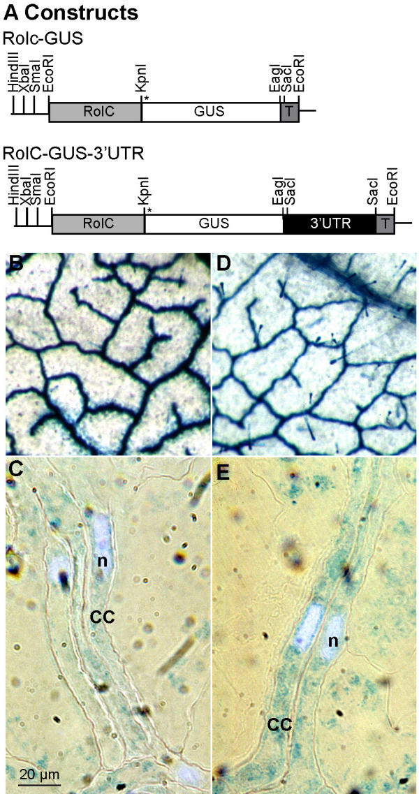
Localization of GUS activity in RolC-GUS transgenic plants. GUS staining was performed on whole tobacco leaves of transgenic plants using X-Gluc as substrate. For higher resolution staining, the leaves were cut to small pieces after the staining, then fixed and embedded in LR White. Thins sections were stained with DAPI for CC nucleus localization and were observed using normal light microscopy coupled to Nomarski imaging and epifluorescence. A) All constructs were prepared in a modified pGPTV-HPT under the control of the CC-specific, RolC, promoter. The asterisk indicates the position of the start codon. T, nos terminator. B) RolC-GUS, C) RolC-GUS-3'-UTR, D) GUS activity localization in leaf thin section of transgenic RolC-GUS, E) GUS activity localization in leaf thin section of transgenic RolC-GUS-3'-UTR. Bar equals to 10 μm.