
Figure 1.
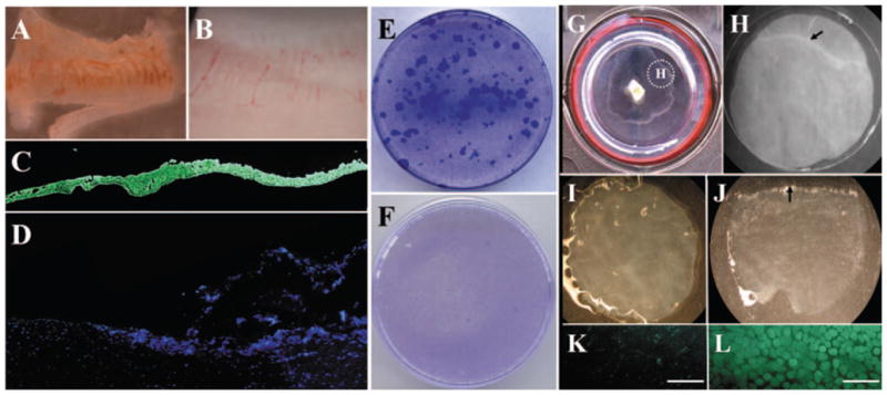
Isolation and clonal culture of human limbal epithelial cells and outgrowth on iAM. From limbal tissue, an epithelial sheet (A) was successfully isolated from the remaining stroma (B) by a modified method of Dispase II digestion. Complete removal of epithelial cells was confirmed by pancytokeratin staining of the isolated epithelial sheet (C), whereas the remaining stroma did not contain pancytokeratin(+) epithelial cells (D). Epithelial cells from the limbal sheet showed vivid clonal growth (E). In contrast, cells isolated from the remaining stroma did not form any epithelial colony on 3T3 fibroblast feeder layers (F). Epithelial outgrowth generated by limbal explant culture on iAM was visible after 2 weeks’ cultivation (G). A 7-mm trephine was used to remove the outgrowth together with the underlying AM (G, inset), and the section was subjected to Dispase II digestion (H, dotted circle in G). The remaining AM (I) did not contain any epithelial cells, as shown by cell-viability assay (K). In contrast, the isolated epithelial sheet (J) contained 100% viable cells, according to the viability assay (L). It should be noted that trephination was intentionally performed to include the border of the outgrowth (H, arrow) and to verify the completion of such isolation by showing such a border of the isolated epithelial sheet (J, arrow). For the data presented in the study, trephination was performed to include only the epithelial outgrowth. Bars, 100 μm.