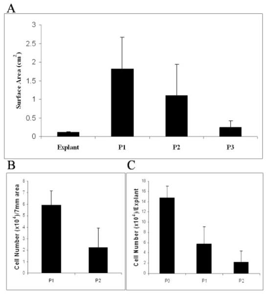
Figure 2.
Decline of limbal epithelial cell outgrowth from limbal explants during successive passages. The surface area of epithelial outgrowth was measured when the same limbal explant was transplanted to a new AM every 2 weeks for three passages. (A) The surface area decreased significantly from P1 to P3 (P = 0.002 for P1 vs. P2 and P < 0.001 for P2 vs. P3; n = 41 for P1, n = 23 for P2, n = 10 for P3). (B) Cells from the outgrowth were isolated and counted, and the number of cells from the unit surface area (7 mm diameter) also significantly decreased from P1 to P2 (P = 0.004, n = 5). (C) The number of cells from the surface of the explant also decreased significantly from P0 to P1 (P = 0.002, n = 5), but did not show a significant difference between P1 and P2 (P = 0.08, n = 5).

