
Figure 3.
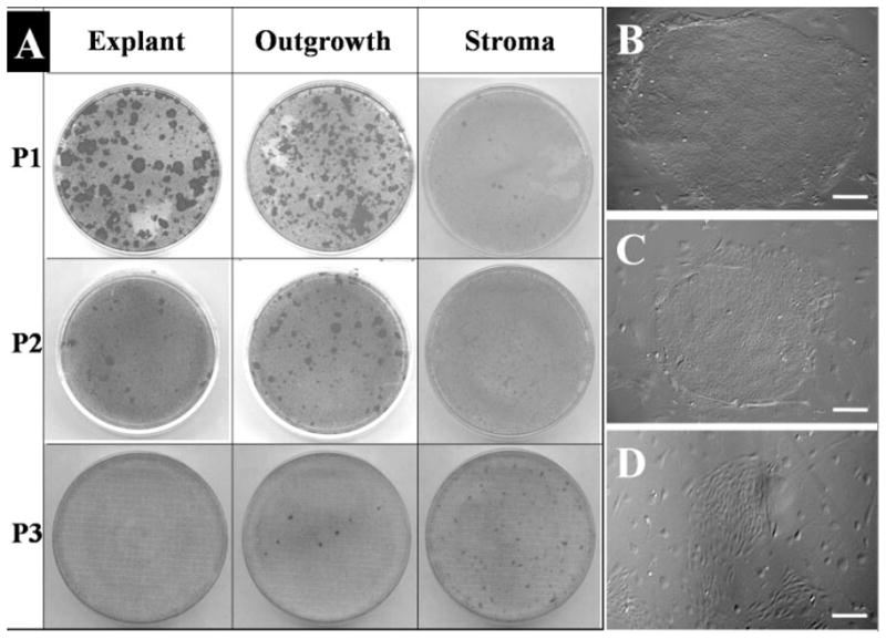
Epithelial clonogenicity of explant surface epithelium, outgrowth epithelium, and remaining explant stroma. The same number of cells isolated from the surface epithelium of the limbal explant and from the explant outgrowth was seeded on 3T3 fibroblast feeder layers for 12 days. Epithelial colonies visualized by crystal violet staining notably decreased from P1 to P3 for both the epithelium on explants (A, left column) and that from the outgrowth (A, middle column). As a comparison, cells isolated from the remaining stroma only yielded increasing fibroblast colonies from P1 to P3 (A, right column). Phase-contrast images showed representative colonies from P1 explant (B), P1 epithelial outgrowth (C), and P3 stroma cells (D). Bars, 200 μm.