
Figure 6.
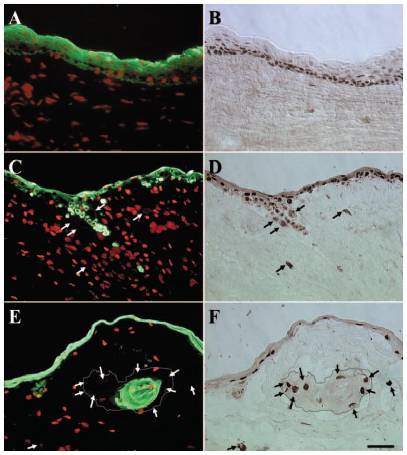
Double staining of pancytokeratin and p63. For P0 explant before cultivation, all cells in the surface epithelium were pancytokeratin(+) (A), whereas most of the basal and some suprabasal epithelial cells showed p63(+) nuclear staining (B). No cell in the limbal stroma showed positive staining for pancytokeratin or p63. In contrast, in P1 and P2 explants, pancytokeratin staining revealed groups of epithelial cells in the stroma (C, E, respectively). In P1 explants, some of these pancytokeratin(+) epithelial cells retained p63(+)nuclear staining (C, D). However, some cells with p63(+) nuclear staining were pancytokeratin(−) (C, D, arrows). Cells showing p63(+) staining but pancytokeratin(−) staining were located at the periphery of the invading epithelial cluster (E, F, arrows, dotted lines outline the border of invading epithelial clusters) for P2 explants. In contrast, cells in the center of invading epithelial clusters were positive for pancytokeratin but negative for p63. Bar, 50 μm.