
Figure 7.
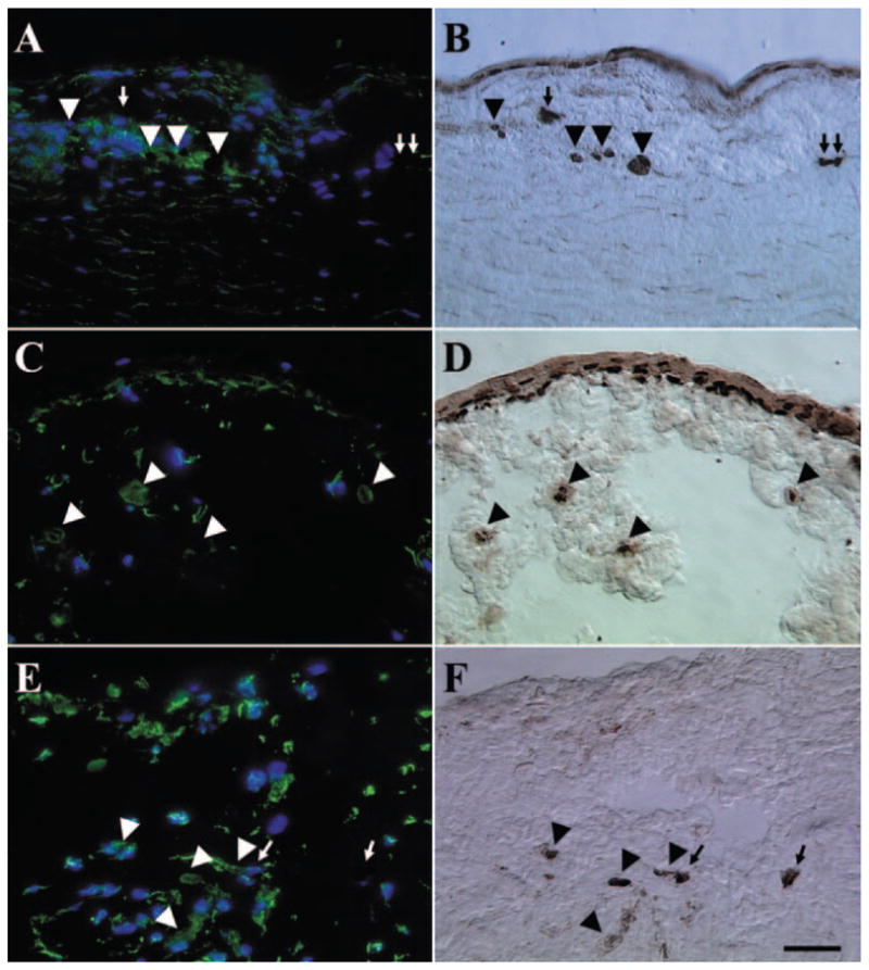
Double staining of vimentin and p63. Vimentin (A, C, E) and p63 (B, D, F) double-stained cells (arrowheads) were found in the limbal stroma of P1 (A, B), P2 (C, D), and P3 (E, F) explants, whereas some p63(+) cells (arrows) did not express vimentin. Vimentin and p63 double-stained cells also presented on the surface of P2 explants (C, D). There was an overall increase of vimentin(+) cells in the limbal stroma from P1 to P3. Bar, 100 μm.